Tuesday, March 11, 2014

chf x ray

Suppose you are looking at a chest x-ray. CHF
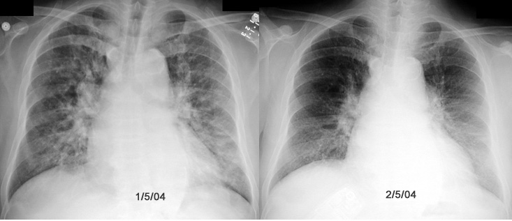
Suppose you are looking at a chest x-ray. CHF

Q2: Anticipated findings in Chest x-ray in a patient with congestive heart

Q2: Anticipated findings in Chest x-ray in a patient with congestive heart

What is the significant x-ray finding on the above film?
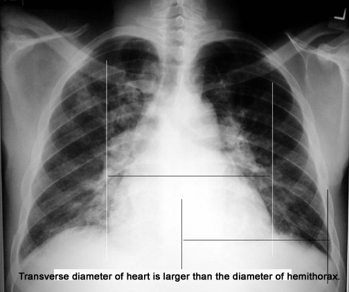
What is the significant x-ray finding on the above film?

Q2: Anticipated findings in Chest x-ray in a patient with congestive heart

Q2: Anticipated findings in Chest x-ray in a patient with congestive heart

Figure 2: Chest X-Ray showing bilateral vascular congestion and cardiomegaly
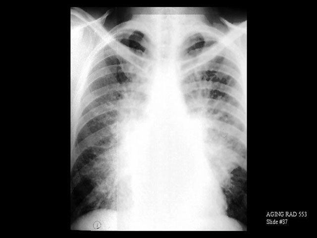
Figure 2: Chest X-Ray showing bilateral vascular congestion and cardiomegaly

X-RAY: PA chest x-ray. Congestive heart failure with interstitial and

X-RAY: PA chest x-ray. Congestive heart failure with interstitial and

The chest x-ray shows blunting of the right costo/phrenic angle and

The chest x-ray shows blunting of the right costo/phrenic angle and

Caption X-ray showing lung congestion due to congestive heart failure.
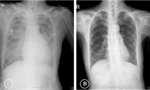
Caption X-ray showing lung congestion due to congestive heart failure.

Figure 1 Chest X-ray showed cardiomegaly with pulmonary edema (A),

Figure 1 Chest X-ray showed cardiomegaly with pulmonary edema (A),

A chest X-ray helps detect problems with your heart, lungs and blood vessels

A chest X-ray helps detect problems with your heart, lungs and blood vessels

 atrial calcification seen in the chest X-ray of a patient with severe MS
atrial calcification seen in the chest X-ray of a patient with severe MS

 Right middle lobe pneumonia; Thyroid goiter; Congestive heart failure

Right middle lobe pneumonia; Thyroid goiter; Congestive heart failure

lateral x-ray of a dog in chf- Suite101.com Images
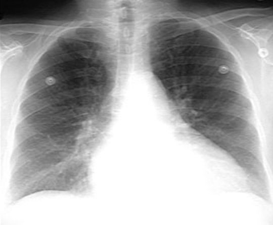
lateral x-ray of a dog in chf- Suite101.com Images

Images of Congestive Heart failure
Images of Congestive Heart failure

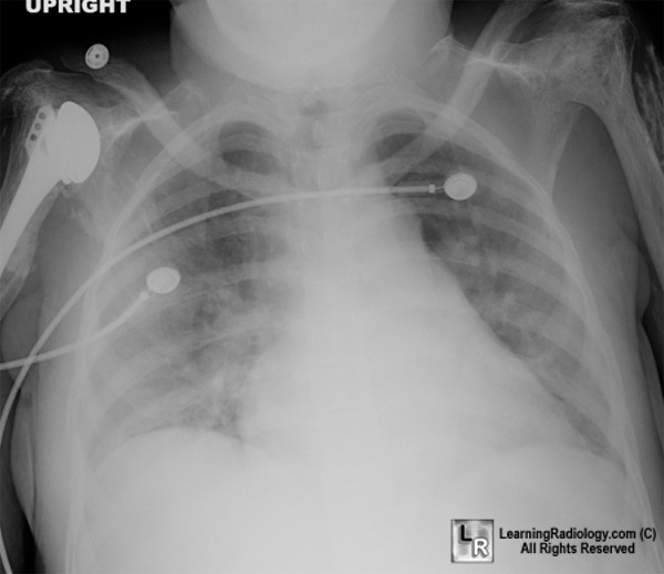
 Acute Chest Syndrome; Dislocated shoulder; Congestive heart failure

Acute Chest Syndrome; Dislocated shoulder; Congestive heart failure

On Day 11, chest X-ray (anteroposterior film) showed progression in right

On Day 11, chest X-ray (anteroposterior film) showed progression in right

Figure 2: Chest X-ray illustrating the cardiomegaly and the broadened

Figure 2: Chest X-ray illustrating the cardiomegaly and the broadened

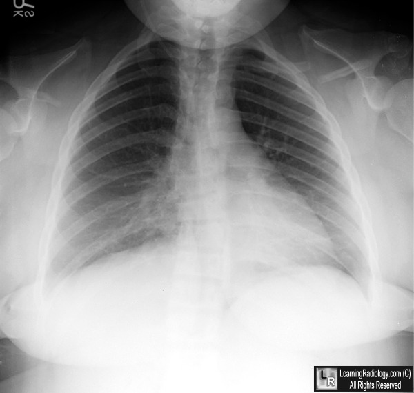
This is a typical chest x-ray of a patient in severe CHF.

This is a typical chest x-ray of a patient in severe CHF.

Chest X-Ray - Heart Failure

Chest X-Ray - Heart Failure

Previous normal chest x-ray (left) and CHF stage II with perihilar haze

Previous normal chest x-ray (left) and CHF stage II with perihilar haze

No comments:

Post a Comment

LinkWithin